








Quick answer: A toothbrush camera can help locate and compare areas that deserve attention. A color, glow, or enlarged image alone cannot identify plaque, stain, food debris, tartar, decay, or a change inside the tooth. Treat every scan as a visual clue, then consider location, cleaning response, symptoms, and changes across repeated scans.
Why Toothbrush Camera Images Can Be Misleading
A close camera view reveals details that a bathroom mirror misses. Extra detail can also create false confidence because light, moisture, angle, and magnification can change surface appearance.
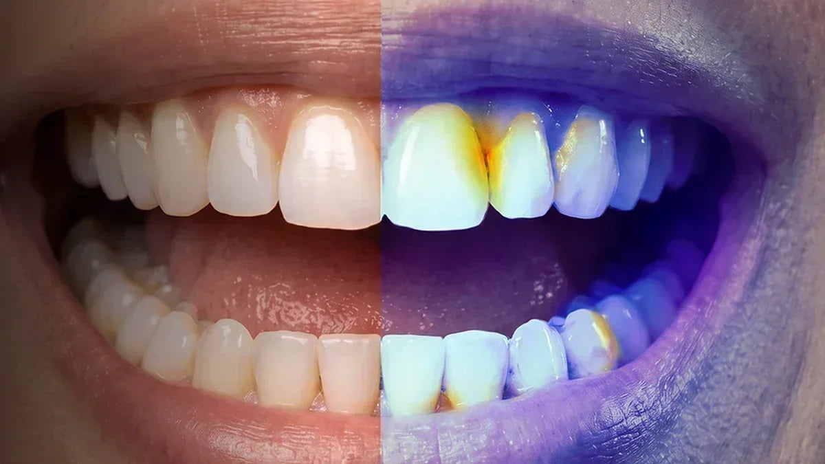
Normal light mainly shows tooth color, surface marks, food, and dental materials. Violet light shows fluorescence, so colors from both modes need separate interpretation. A dark mark under normal light may not appear under violet light. A fluorescent area may look ordinary under normal light.
Saliva, water droplets, reflections, shadows, and close magnification can sharpen edges or deepen colors. Distance, camera angle, room light, and tooth dryness also reduce scan-to-scan consistency. A camera supports observation, but cannot provide a dental diagnosis.
Plaque vs. Food Debris vs. Stains vs. Tartar
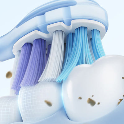
Several common findings can look similar on a small screen. Location, texture, movement, and response to rinsing or cleaning offer more useful clues than color alone.
| Check | Plaque | Food debris | Stain | Tartar |
|---|---|---|---|---|
| Common areas | Gumline, gaps, back teeth | Gaps and molar grooves | Outer enamel surfaces | Gumline and inner lower front teeth |
| Typical appearance | Often colorless, soft, sticky, or fuzzy | Irregular shape; color may match recent food | Flat color that stays in a stable area | Hard, fixed deposit; often yellow or brown |
| After rinsing | Usually remains | May move or disappear | Usually unchanged | Unchanged |
| After brushing | May decrease | Often clears | May remain | Remains |
| Professional care | Useful for persistent buildup or gum symptoms | Useful for repeated food trapping | Useful for sudden color change or symptoms | Required for removal |
Cleveland Clinic describes plaque as a sticky, colorless film and tartar as hardened buildup that normal brushing and flossing cannot remove. The table provides observation clues, not confirmation. Never use a sharp object to scrape a suspected deposit.
What Red or Pink Areas May Show on the D50
Violet-light images focus on fluorescence rather than natural tooth shade. A red or pink area can point toward plaque-related compounds, yet signal strength varies across people and tooth surfaces.
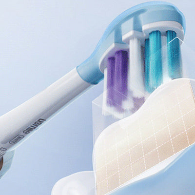
The usmile D50 OralCam Toothbrush uses 405 nm violet light and QLF technology. Certain compounds associated with plaque bacteria can give off red or pink fluorescence. “Plaque-related signal” is a more accurate label than “confirmed plaque.”
Clinical research links stronger red fluorescence with more mature biofilm. Another clinical study found meaningful variation across tooth sites and individuals. Brightness cannot grade cavity severity, gum disease, or overall danger. No visible highlight also fails to prove a plaque-free surface. Fresh or thin plaque may appear faint.

Location Matters: Gumline, Gaps, Molars, and Dental Work
Color becomes more useful after location enters the picture. A gumline signal calls for a different response from material caught in a molar groove or beside a crown.
- Gumline: Plaque often collects along the edge where teeth meet the gums. Also note bleeding, swelling, tenderness, or recession.
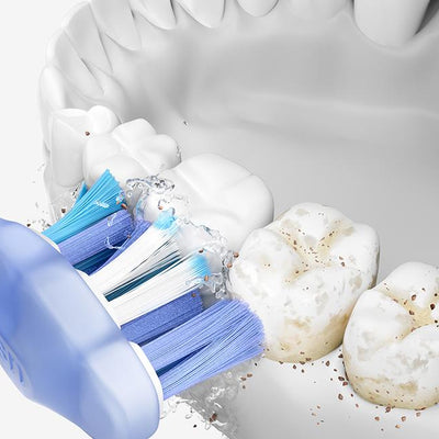
- Gaps: Food debris is common between teeth. A toothbrush may miss material that floss or an interdental cleaner can reach.
- Molar grooves: Deep grooves can hold food and stain. Shadows and natural tooth anatomy can also look like dark pits.
- Fillings, crowns, and braces: Dental materials can reflect light or show a different color from natural enamel.
Repeated food trapping, unpleasant odor, soreness, or a persistent change in the same area deserves dental attention.
The Rinse, Clean, Brush, and Rescan Check
A short and repeatable check can help separate loose material from a fixed change. Similar camera conditions give each comparison greater value and reduce confusion from light or angle.
- Record the location, color, shape, and size of the area.
- Rinse with water, then scan from a similar angle.
- For a spot between teeth, use floss or a suitable interdental cleaner.
- Follow D50 zone prompts and brush with normal pressure.
- Rescan from a similar distance, angle, and light level.
- Compare both scans along with any pain, bleeding, odor, or sensitivity.
If the Area Disappears or Moves
Food debris or a loose surface deposit becomes more likely. Repeated trapping in the same gap can still signal a contact, gum, or restoration issue that needs professional review.
If the Plaque-Related Signal Becomes Weaker
Cleaning created a visible change. The result supports better removal in that area, but cannot prove that every layer of plaque has gone.
If the Area Stays in the Same Place
A stain, tartar deposit, filling material, natural tooth feature, or tooth change remains possible. Avoid aggressive scrubbing, sharp tools, or strong whitening products chosen from camera color alone.

Use the D50 to Track Cleaning Patterns, Not Diagnose Conditions
Repeated scans from the same person offer greater value than a dramatic single image. Consistent views can reveal areas that often need extra cleaning attention.
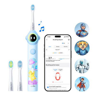
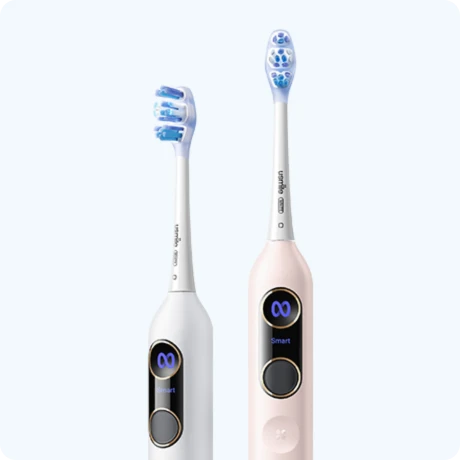
D50 combines a camera toothbrush design with smart electric toothbrush features. It displays scan results on the handle, provides zone prompts, adjusts brushing intensity near plaque-related areas, and syncs progress with the usmile App. Early use can include scans around brushing to create a personal baseline. After results remain low and stable, weekly trend checks may be enough, following official D50 advice.
For readers comparing electric toothbrushes for plaque removal, the useful question is not which model shows the brightest color, but whether routine cleaning improves the same areas over time. An electric toothbrush with app support can help document those patterns, but it cannot diagnose disease.
Compare the same tooth position under similar conditions. Another person’s color or brightness offers little value because oral bacteria, enamel, dental work, moisture, and camera technique vary. Personal change over time gives a clearer signal.

When a Toothbrush Camera Is Not Enough
Some findings need a clinical examination rather than more camera checks. Symptoms, hard deposits, and persistent color changes can involve structures that a home image cannot evaluate.
Contact a dentist for:
- Persistent pain, cold sensitivity, heat sensitivity, or pain during biting
- Gums that keep bleeding, swelling, or pulling away from teeth
- A visible hole, crack, dark pit, or black spot
- A single tooth that suddenly becomes gray or much darker
- A fixed, hard deposit that normal brushing cannot remove
- Repeated food trapping, unpleasant odor, or discomfort in the same area
- A suspicious area that remains unchanged after several days of normal care
More scans cannot replace an examination, dental X-rays, professional cleaning, or a periodontal assessment.
Frequently Asked Questions About Toothbrush Camera Images
Unfamiliar colors often raise the same practical questions. The short answers below separate useful home observations from findings that still need professional examination.
Does no red fluorescence prove there is no plaque?
No. Fresh or thin plaque may show little fluorescence. Distance, angle, moisture, and light can also hide a weak signal.
What does plaque look and feel like on teeth?
Plaque is often almost colorless. A tooth surface may feel sticky, rough, or fuzzy, especially near the gumline.
Does all plaque glow red under violet light?
No. Biofilm age, tooth surface, bacterial composition, camera conditions, and personal variation can affect fluorescence.
Does brighter red signal more dangerous plaque?
Not necessarily. Stronger red fluorescence can relate to mature biofilm, but brightness cannot grade cavity risk or gum disease severity.
Can food debris look like plaque on a camera?
Yes. Food can create plaque-like shapes in gaps and molar grooves. Rinsing, interdental cleaning, and rescanning can reveal movement or disappearance.
Can tooth stains fluoresce or look like plaque?
Yes. Stains, dental materials, shadows, saliva, and reflections can create similar colors. Color alone cannot confirm the cause.
Can the D50 detect tartar?
D50 may reveal a fixed deposit, but a camera image cannot confirm tartar. Hardened buildup needs professional removal.
Can a toothbrush camera detect cavities?
No home camera can diagnose decay from color alone. A dark pit, visible hole, pain, or sensitivity needs a dental examination.
Why can the same area look different between scans?
Camera angle, distance, tooth moisture, saliva, reflections, and room light can alter color, contrast, and edge sharpness.
How often should I use the D50 checkup feature?
Daily scans around brushing can help establish a personal baseline at first. Weekly checks may be enough after scans consistently show low buildup.
Can D50 replace dental checkups?
No. D50 supports daily cleaning and trend tracking. Dental visits assess decay, gum health, tartar, restorations, and problems hidden from a camera.